The ICL Surgery Procedure
Implantable Collamer Lenses (ICL) are suitable for patients between the ages of 18 and 50, with high-degree refractive errors (high myopia, hyperopia, and high astigmatism). The ICL surgery procedure does not change the structure of the eye, which means that any time and for any reason, the lenses can be removed and the eye can easily go back to the state before the surgery. Unlike the ICL procedure, with LASIK intervention a small part of the cornea is permanently removed; with RLA (refractive lens exchange), the natural lens is replaced with an artificial lens (an implant).

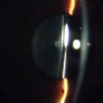
The ICL surgery is executed in the following way: an extremely thin implant is placed between a person’s iris and lens, thus fully correcting a refractive error and enabling excellent visual acuity. This way it is possible to correct myopia to -23 diopter, hyperopia to +12 diopter, and astigmatism to 6 diopters. Unlike multifocal implants, this type of surgery does not require removal of the person’s lens, and the placed implant stays in the eye permanently. After the surgery, there is no need to use glasses or lenses anymore. However, this procedure cannot replace hyperopia glasses for presbyopia.
The ICL is inserted in the posterior chamber of the eye, directly next to the person’s lens. These implants are produced individually for every patient because every eye is specific and requires optimal correction and custom-made implants. The material of the implant is made of recombinant collagen and contains a UV filter that protects the person’s lens from UV damage.
A course of Intervention:
- Anesthetic – The eye is anesthetized with eye drops.
- The first step before the intervention – at the edge of the cornea a minimal incision is made, about 3 mm in diameter, through which the ICL is implanted.
- Implanting and positioning of the ICL – a small soft lens (implant) rolled like a scroll is implanted into the eye through a small opening with a help of a special injector, and then smoothed out into the place.
- The intervention is finished. Most patients immediately notice a significant improvement in their vision.
This is a minimally invasive procedure that lasts about 10 minutes per eye. Usually, both eyes are operated on within a week.
Postoperative Follow-up
After the surgery, the operated eye is closed with a “cover shell”, which is taken off the next day after the follow-up examination and detailed information on therapy. A couple of days after the intervention the vision is slightly blurry, but the visual acuity gradually improves. The first few days after the surgery it is necessary that you follow the instructions, shield your eye before sleep, and avoid physical exertion.